X-ray Vs CT Scan Vs MRI
Protocols
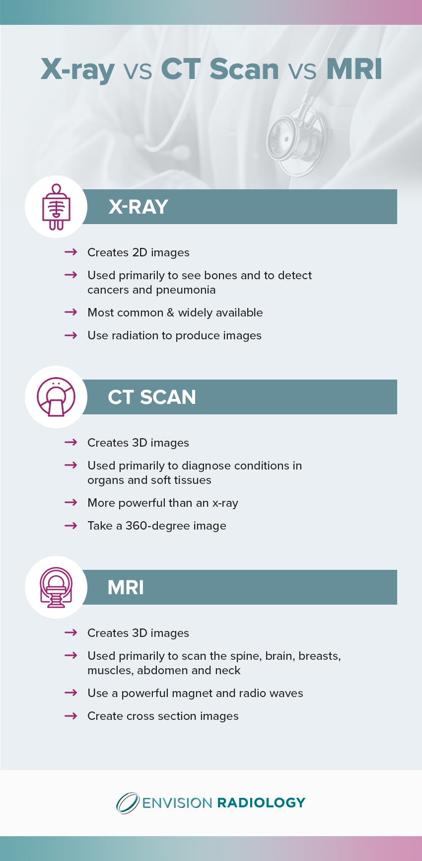
X-ray Vs CT Vs MRI
What Is an X-Ray?
X-rays are the most used diagnostic imaging test and are widely available. Even if you requiremore sophisticated body scans, it’s likely you’ll receive an x-ray first.
They are a form of radiation, and when passing through your body, bone and other dense objects block the radiation and look white on the film of the x-ray. The less dense tissues are hard to see and appear gray. While limited exposure to radiation from x-rays isn’t harmful, if you’re pregnant, the doctor will take special precautions.
The doctor will position the part of your body for scanning between the digital x-ray sensor or photographic film and x-ray machine. While the machine sends the radiation briefly through your body, you need to stay still.

Types of X-Rays
There are two primary types
of x-rays — soft and hard.
1. Soft x-rays have fairly short
wavelengths of approximately 10 nanometers (nanometers are one-billionth of a
meter). Therefore they can be placed in the electromagnetic (EM) spectrum
between gamma-rays and ultraviolet (UV) light.
2. Hard x-rays have wavelengths of
approximately 100 picometers (picometers are one-trillionth of a meter)
wavelengths. They occupy the same area as gamma-rays on the EM spectrum.
What Is a CT Scan?
A CT scan generates high-quality, detailed images of
the body. It’s a more powerful and sophisticated x-ray that takes a 360-degree
image of the spine, vertebrae and internal organs. You may have a contrast dye
injected into your blood so the doctor can see your body structures more
clearly on the CT scan.
1. Positron Emission Tomography (PET)
CT Scan
The PET CT scan helps the physician
to see the level of activity of certain body organs and tissues, along with
their structure. You’ll receive a substance called a “tracer” containing
glucose with a little bit of radioactive material attached before your test.
This tracer travels through your body
systems. It acts like a dye for the imaging scan to pick up on. If there’s high
chemical activity in certain areas, more of the dye will be picked up, and it
will show bright spots on the image, alerting the doctor of possible disease.
The radiation dose in the tracer is
safe and minimal for most individuals. The tracer will be swallowed, inhaled or
injected, depending on the examined body part.
Physicians use PET scans often for
detecting heart problems, cancer and brain diseases.
2. CT Urography
CT urography is a type of specialized
radiological exam used for evaluating the urinary tract, which includes the
ureters, kidneys and bladder. It’s an innovative technology that uses computed
tomography to produce cross-sectional images throughout your body. The images
of internal organs are very detailed and allow doctors to make decisions on the
most accurate treatment plan to take.
The most common uses for this exam
are to evaluate blood in your urine and detect kidney stones.
What Is an MRI?
MRI stands for magnetic resonance
imaging and combines a strong magnet with radio waves. A computer operates the
magnetic components, creating incredibly detailed images of body structures.
The doctor views the images as “slices” or cross-sections of the scanned body
part. Unlike x-rays, there’s no radiation involved. Doctors use MRI scans
frequently for diagnosing joint and bone problems, as well as for assessing
treatment progress, looking into brain abnormalities and evaluating pelvic pain
or infertility issues.
1. Open MRI Scans – High-Field
(1.5T)
A high-field (1.5T),
open MRI delivers superb image quality:
- Open
MRI: This refers to the configuration of the
equipment. The Open MRI creates a bright imaging and a relatively roomy
experience because it is open on three sides. It has a modern design which
yields quick and comfortable exams, which is particularly beneficial for
individuals who are claustrophobic.
- High-Field
(1.5T): High-field (1.5T) is referring to
image quality. The 1.5T offers a wide range of coil options, allowing for
better image quality across some imaging applications when compared to the
3T.
2. MRI Short Bore Scans –
High-Field (1.5T)
The bore refers to the opening in the
MRI imaging machine. Short bore MRI scans are
5 percent wider and 50 percent shorter than the conventional MRI setup. The
dimensions provide the patient with a roomy and very airy imaging experience.
3. MRI Open Bore Scans –
High-Field (1.5T)
An open bore MRI provides
a wider opening that allows for a far more comfortable MRI scan. With the
traditional bore, there’s a slightly bigger opening than the patient, and this
creates a very uncomfortable, restrictive environment.
4. MRI Scans – High-Field (1.5T)
This high-field (1.5T) MRI scanning
machine offers the most innovative imaging technology. It’s valuable for
doctors to scan all body parts and is considered the industry standard.
5. 3T MRI Scans
Also known as the 3 Tesla MRI, the 3T MRI scan is
an efficient and powerful imaging exam that you may have instead of the 1.5T
traditional scan. While 3T scanners were once only found mainly in medical
research centers, these days, you may see them in clinical settings too.
The 3T scans use strong, powerful
magnets, producing a magnetic field much more powerful than the 1.5T scan. This
allows the MRI to create clearer images more quickly.
6. MRI Spectroscopy
MRI spectroscopy is a non-invasive method
used for characterizing the biochemistry of infarcts, tumors and other
pathology. It’s frequently performed to diagnose specific metabolic disorders
such as those that affect the brain. It helps doctors figure out the specifics
of tumors like their metabolism or aggressiveness.
7. MRCP Scans
Magnetic resonance
cholangiopancreatography (MRCP) scan is a specific type of MRI that focuses on
attaining images of the pancreatic and hepatobiliary systems, including the
liver, gallbladder, pancreas, bile ducts and pancreatic duct.
Differences between CT scan and MRI
| CT scan | MRI |
|---|---|
| Radio-frequency wave is used | |
| No radiation hazard | |
| Multiplanner images are produced | |
| Soft tissue discrimination is excellent | |
| Calcification appears as signal void, so not sensitive, only bone marrow changes are identified | |
| No known hazard in pregnancy | |
| Contraindicated if metallic prosthesis is present (except for MRI-friendly prosthesis) | |
| Not suitable for acute case due to motion artefact | |
| White matter diseases are better evaluated | |
| More expensive than CT | |
| Specialized cooling system is required, especially in magnet superconductor | |
| Claustrophobic effect present | |
| More time consuming |