MRI Basic
Protocols
MRI scan
MRI is a nonionizing imaging technique using magnetic fields and radio-frequency
waves to visualize the anatomical structures.
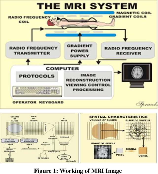
Basic principle
Magnetic resonance imaging depends on the magnetic properties of the nuclei of certain elements.
In order to perform MRI, we first need a strong magnetic field. The field strength of the magnets
used for MR is measured in units of Tesla. For medical purpose, MRI is based on hydrogen nuclei or
protons in water and lipids. Each proton can be considered to be a spinning positive charge with an
associated magnetic field. Thus, a proton is said to have a magnetic moment. Bringing tissue in
an external magnetic field results in an alignment of these tiny magnetic moments either parallel or
anti parallel to the applied field direction. Now a radiofrequency pulse at an appropriate frequency
(resonant frequency) is applied and a proportion of the protons change their alignment. After this, the
protons return to their original position. As they realign (relax), they produce a radio signal that can be
detected by coils placed around the patient. This signal depends on the density of protons and on two
relaxation times T1 and T2. T1 represents the time protons take to return to alignment, and T2 represents
the time they take to diphase. This information is then analysed by a computer which generates an image
representing the distribution of protons in that part of the body.

Advantages:
- • MRI has not only established itself as a powerful tool for fast routine scanning but also expanded its
- application into studying organ function, metabolism and physiology.
- • The use of nonionizing radiation. Therefore, free from any biological hazards.
- • Direct multiplanner imaging in transverse, coronal and sagittal planes or in any other desired plane.
- • MRI images of the brain and other cranial structures are more clear and detailed than with other imaging methods.
- This detail makes MRI an invaluable tool in the early diagnosis and evaluation of many conditions, including tumour.
- • MRI enables the detection of abnormalities that might be obscured by bone with the other imaging method.
- • Contrast material used in MRI is less likely to produce an allergic reaction than the iodine-based
- materials used for conventional X-ray and CT scanning.
- • MRI is the most sensitive investigation for brain tumour.
Disadvantages:

Contrast CT scan and MRI
Although CT scan and MRI can provide valuable information about the anatomy and pathology of internal
organs and tissues, greater details can be obtained by adding a contrast. It may help visualize the pathology
more clearly and indicate the nature of the lesion by the amount and distribution of contrast uptake. This is a
useful add-on to the conventional CT scan and MRI, and is used in about 50% cases.
Iodine-based contrast agents such as Iopamiro 300 are used with CT scan. It is given intravenously prior to scanning.
Owing to the high atomic number of these agents, it can attenuate the X-ray beams and appear brighter or
radio-opaque in the film. Blood containing this contrast agent flows through the vessels of the area to be
scanned. On the film, the agent increases the contrast between blood and surrounding tissues. This facilitates
visualization of the arteries, veins and ultimately the internal structures. Highly vascularized areas will
become brighter on the film.
In addition to the intravenous contrast, oral radio-opaque agents (barium) can be used for contrast CT
scan of abdomen. The liquid should be taken several hours prior to the scan.
Adverse effects of contrast agent used in CT scan include severe and potentially life-threatening
allergic reaction. It may induce kidney damage with increased risk in patients with pre-existing
renal insufficiency, diabetes or reduced intravascular volume. Patients with mild renal impairment should
maintain full hydration for several hours before and after the injection. But for the moderate renal failure,
iodinated contrast should be avoided. Patients undergoing dialysis may safely undergo contrast CT scan.
In MRI, contrast agents are usually selected for their specific magnetic properties. Most commonly, a paramagnetic
contrast agent like Gadolinium is used. This is given intravenously. Tissues that are enhanced by the contrast appear
very bright on T1-weighted image. It increases the contrast between areas with high blood flow and that with low blood flow,
and thus facilitates better visualization. This is helpful in the detection of primary and secondary tumours, abscess, infection
and inflammation. This also helps to measure the size of a lesion (especially tumour) by delineating its border. It is an important
tool for follow-up after chemotherapy or radiotherapy since the changes in blood flow, revealed by contrast MRI, may indicate
the result of the therapy. This is also useful in assessing tissue damage and perfusion in stroke. Unfortunately, Gadolinium is
considered toxic in patients with renal failure. Therefore, patients should be screened for renal function prior to scanning.
Haemodialysis following a contrast MRI scan is an option for such patients.
Superparamagnetic contrast agent, such as iron oxide nanoparticles, has been recently developed for the contrast MRI imaging
of liver and abdomen. These agents appear very dark on T2-weighted films and accumulate in the normal liver, but not in abnormal areas.
When taken orally, these agents help to visualize the gastrointestinal tract (GIT). Diamagnetic agent, such as barium sulphate,
is less frequently used in MRI of the GIT.