USG of Pregnancy Profile(P/P)
Protocols
Early Pregnancy usg(upto 13 weeks)
☯️Condition:
প্রস্রাব এর চাপ থাকতে হবে।

☯️Parameters :
1️⃣Gestational Sac(GS): সাধারণত ৫ সপ্তাহে পাওয়া যায়।
2️⃣Cardiac activists : সাধারণত ৮ সপ্তাহে পাওয়া যায়।
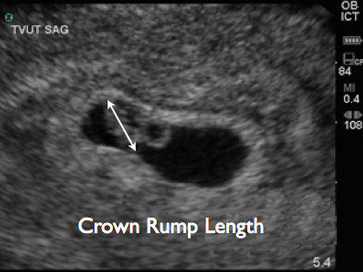
3️⃣ Crown rump length(CRL): More accurate.
(Length from head to buttock)
সাধারণত ৭ সপ্তাহে মাপা যায়।
4️⃣Yolk Sac: সাধারণত ৬ সপ্তাহে পাওয়া যায়।
🚼Fetal Heart Rate: FHR সাধারনত ৮ সপ্তাহে পাওয়া যায়। usg mechine এ M-mode এ count করা হয়।
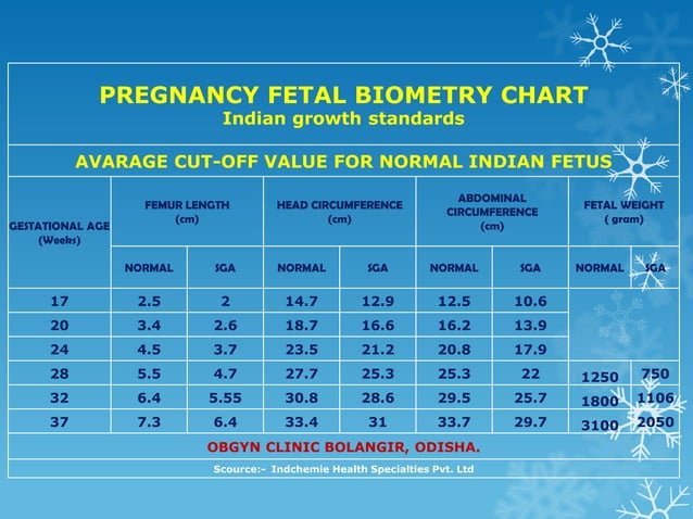
Fetal Biometry: Parameters:


Fig: Femural length
🚼Bi-parietal diameter (BPD) :নিচের criteria গুলো আমরা usg তে পাবো। অবশ্যই একটি oval picture না আসলে head এর measure নিবো না, তাহলে কম বা বেশি EDD আসতে পারে।
Head Looks like:
➡️head should look oval
➡️Symetrical skull
➡️Both thalami should be visible
➡️Interhemisphereic fissure / Falx cerebri should be visible.
Measure from outer table to inner table of skull.
🚼Femoral length(FL):
➡️The entire femur should be imaged but the bright
reflectionof the cartilaginous epiphysis should not be
included.
🚼SN:
➡️BPD & FL এর ব্যবধান সাধারনত ১৫ দিনের বেশি হবে না। ১৫ দিনের বেশি হলে আবার রিপিট করে দেখতে হবে।
➡️যারা মাত্র usg শুরু করছেন তাদের জন্য FL clear আনা একটু কষ্ট কর। যদি কেউ FL না আনতে পারেন তাহলে শুধু ভালো করে BPD
এর মাপ নিয়ে EDD দিতে পারেন। আর দুটোই আনতে পারলে average date টা দিবেন।
অনেক usg mechine একাই average date Count করে দেয়।
➡️usg যারা প্রথম প্রথম করবে, তাদের প্রতি উপদেশ তারা হুড়ো করবেন না, ভয়ও পাবেন না। ভালো করে history নিবেন।
🚼Abdominal Circumference(AC):এর দুইটা variety আছে Elliptical & Vertical measures.
➡️Elliptical টা দিয়ে আমরা বাচ্চার wt দেখতে পারে। পাশের ছবিতে সুন্দর করে দেওয়া আছে।
এটা দিয়ে বাচ্চার wt measures করা হয়।

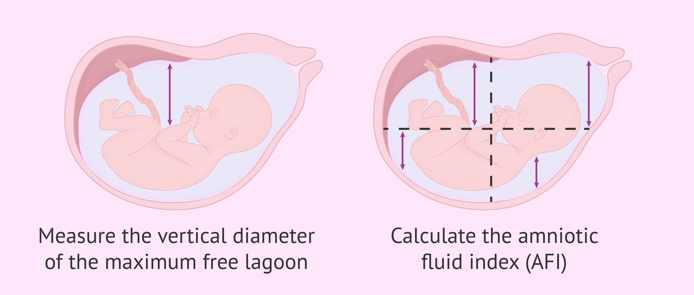
Fig: AFI
🚼amniotic Fluid :AFI
➡️Normal range: 10-20 cm (Ref: Rumack); But INM follow 8-24 cm.
➡️How to measure:পাশে ছবি দেওয়া আছে।
1.Divide the abdomen into 4 quadrants by an immaginary line passing vertically & horizontally
through umbilicus.
2. Then place the probe vertically &
measure the maximum vertical length of amniotic fluid in
every quadrant.AFlis the summationof all four maximum
3.vertical lengths.N.B- No pockets should contain any fetalpartor
umbilical cord & the pocket should be at-least 1cm wide.
Molar Pregnancy:


1.Enlarge uterus containg echogenic tissue that expands the endometrial canal with innumerable ,diffusely & uniformly
distrubuted cystic spaces,ranging in size from 2-3 cm (Hydropic villi)
2.Solid collection of echoes with multiple anechoic spaces too many
in number .Typical appearance of branch of grapes.
Blighted ovum /Anembryonic gestation:

if gestational sac size is >25 mm but inside of gestational
sac there is no yolk or embryo.
Anatomy:

Amniotic Fluid Index(AFI):
Normal range: 10-20 cm 0r 8-24 cm

Procedure:
1. Divided the abdomen into 4 quadrants by an immaginary line
passing vertically & horizontally through umbilicus.
2. then place the usg probe vertically & measure the maximum vertical
length of amniotic fluid in every quadrants.
3. AFI IS the summation of all four maximum vertical length.
(No pokets should contain any fetal parts ,umlical cord & pocket should at least 1 cm)


Placental Maturity: Grading
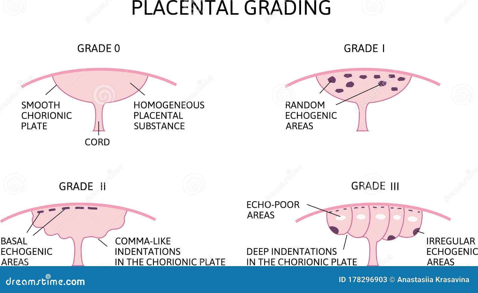

Grade-0: No visible calcification & smmoth chronic plate ( 0- < 18 wks)
Grade-1: Scattered tiny calcification & subtle indentations of chronic plate ( 18- <30 wks)
Grade-2: (30 - < 39wks)
1.larger basal & comma like echo
2.Large indentation of chronic plate
Grade-3 : ( > 39WKS)
1. Extensive basal echogenicity and circular echodensities fully outlies cotyledoms.
2. complete indentation of chronic plate
Usg of placenta Praevia:

Types:
1.Lowlying
2.Marginal
3.partial
4.complete placental praevia
USG Findings:
Presence of echolucent or circular lines overlying the internal os on transabdominal or
transvaginal ultrasound should alert the examiner to the presence of vasa previa.
This can be confirmed by transvaginal assessment with color and spectral Doppler,
confirming presence of arterial fetal vessels
